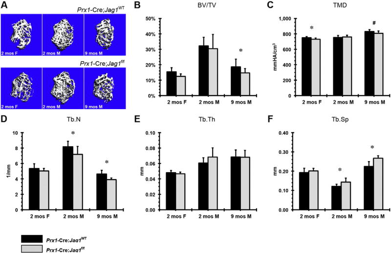
Fig. 3.
µCT analysis of trabecular bone in Prx1-Cre;Jag1f/f mice at 2 months (females and males) and 9 months (males) of age: (A) representative images, (B) bone volume fraction (BV/TV), (C) tissue mineral density (TMD), (D) trabecular number (Tb.N), (E) trabecular thickness (Tb.Th) and (F) trabecular separation (Tb.Sp) (*p < 0.050, #p < 0.100).

