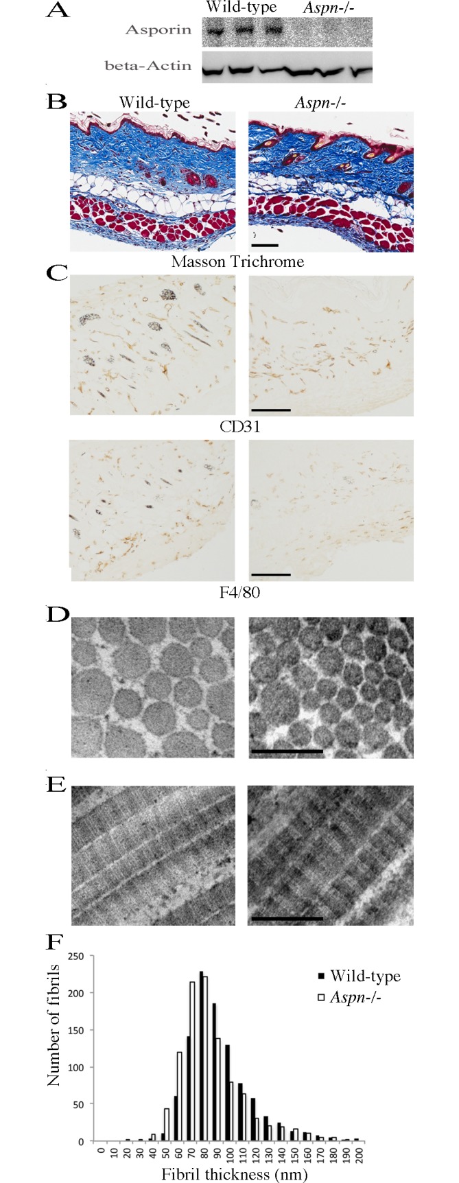
Fig 2. Skin phenotype of Aspn-/- mice.

(A) Asporin immunoblotting of dorsal skin extracts from three wild-type and three Aspn-/- mice. (B) 2 month-old female dorsal skins were analyzed histologically by staining with Massson Trichrome, or (C) by immunohistochemistry by staining for the presence of CD31-positive blood vessels and F4/80-positive macrophages; bars in A and B are 100 μm. Similar results were obtained from analyses of six mice from each genotype. (D) Transmission electron microscopy on cross-sectioned collagen fibrils in reticular dermis. (E) Transmission electron microscopy on longitudinally oriented collagen fibrils in reticular dermis. Bars in C and D are 200 nm. (F) Quantification of collagen fibril diameter in reticular dermis. 1,000 fibrils were measured from electron microscopy images collected from six wild-type and six Aspn-/- mice.
