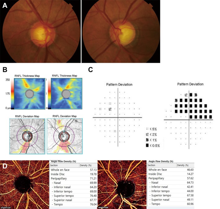
Fig 2. A representative case of unilateral PG eye (left eye) and fellow PPG eye (right eye).
The patient was a 64-year-old man who was referred to the glaucoma clinic on annual examination. Treated intraocular pressure was 14 mmHg for both eyes. (A, B) Color disc photography and retinal nerve fiber layer (RNFL) thickness map and deviation map on spectral-domain OCT showing inferotemporal RNFL defect with optic disc hemorrhage in the right eye and notching in the left eye. (C) Visual field tests showing superior arcuate scotoma in the left eye. The right eye showed no abnormality. (D) Angioflow views are acquired from internal limiting membrane to RNFL posterior boundary. Based on gross observation, the right eye showed no drop-out of peripapillary vasculatures. However, the left eye showed wedge-shaped focal vascular reduction in the same area as RNFL defect. On angioflow density, inferotemporal vessel density in right eye was higher than that in left eye (69.00% versus 44.00%).

