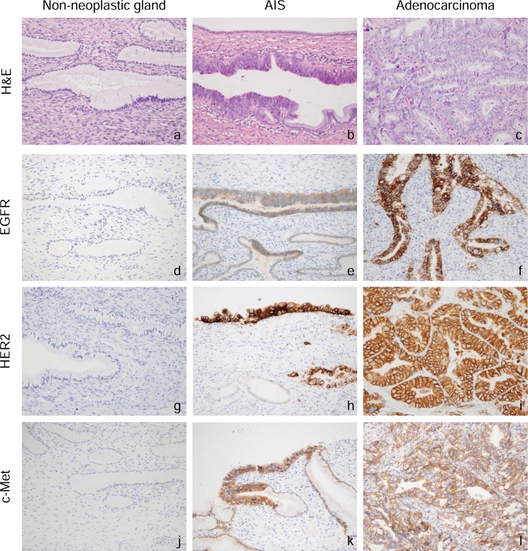
Fig 1. H&E staining and immunohistochemical staining in surgical specimens of non-neoplastic cervical glands, adenocarcinoma in situ (AIS), and cervical adenocarcinoma.
Representative immunohistochemical staining of EGFR (d-f), HER2 (g-i), and c-Met (j–l) is shown. The receptor tyrosine kinases were predominantly expressed on the membranes of tumor cells.

