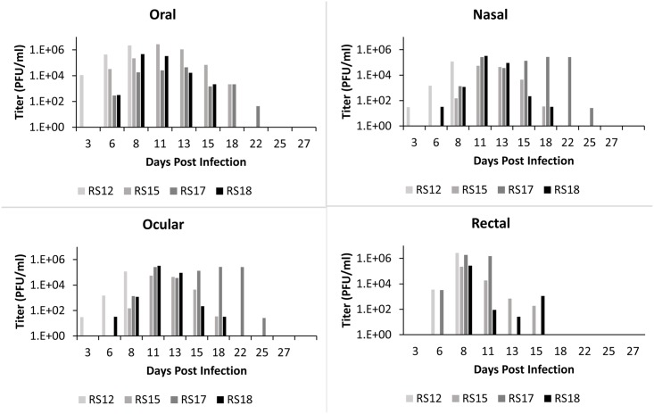
Fig 8. Shedding of MPXV in African rope squirrels infected intradermally with MPXV-Congo/Luc+.
Swabs were taken from oral, ocular, nasal and rectal mucosa then tested by TCID50 assay to determine the concentration of virus shed in PFU/mL. Titers increased following infection, peaked between days 8 and 13, and then decreased for all animals except RS17, which showed consistently high titer from ocular swabs until day 25 pi.

