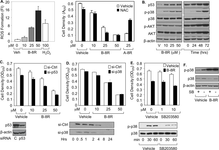
Fig 5. Peptide B-8R induces elevated ROS, p53, and activated p38 in prostate cancer cells.
(A, B) C81 cells were treated with Vehicle, different μMolar concentrations of Peptide B-8R, or A-8R without or with 0–5 mM NAC (N-acetyl cysteine) and monitored for (A) ROS generation after 0–30 min as measured by fluorescence intensity (FI) or (B) cell number after 3 days of incubation as measured by MTT assay. (B) C81 cells were treated with Vehicle or different concentrations of Peptide B-8R, as shown, or 25 μM Peptide B-8R for 0–72 hrs and monitored for by Western blotting for expression of p53, P-p38, p38, p-AKT, or AKT. C81 cells were treated with different concentrations of Peptide B-8R, as shown, in the presence of (C) transfected p53 or Control (Ctrl) siRNA, (D) transfected p38 or Control (Ctrl) siRNA, or (E) Vehicle or 10 μM SB203580 and Western blotting is shown below each figure for p53, p-38, p-p38. Bar graphs represent averages of three independent experiments plus standard deviations. Asterisks indicate statistical significance (P<0.04) of (A) ROS Formation of Peptide B-8R or H2O2 relative to Vehicle or cell density of NAC-treated cells relative to Vehicle or (C) cell density of cells treated with si-p53 relative to si-Ctrl. (F) C81 cells were treated with Vehicle or 25 μM Peptide B-8R, in the absence or presence, as shown, 10 μM SB203580 (SB) and Western blotting was used to measure p-38 and p-p38. Note that β-actin was used to control for protein loading.

