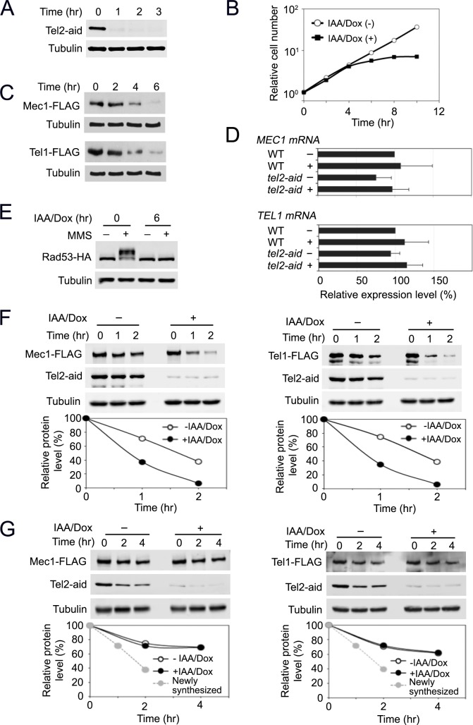
Fig 1. Effect of Tel2 depletion on Mec1 and Tel1 functions.
(A) Expression of Tel2-aid after AID activation. Cultures of tel2-aid cells were treated with 3-Indoleacetic acid (IAA) and doxycycline (Dox) for the indicated time periods. Cells were analyzed by immunoblotting with anti-AID or anti-tubulin antibodies. (B) Cell proliferation after Tel2 depletion. Cultures of tel2-aid cells were treated as in A. Cells were counted using a hematocytometer under a microscope. (C) Expression levels of endogenous Mec1 or Tel1 protein after Tel2 depletion. tel2-aid cells expressing Mec1-FLAG or Tel1-FLAG were incubated with IAA and Dox for the indicated time periods. Cells were subjected to immunoblotting analysis with anti-FLAG or tubulin antibodies. (D) Levels of MEC1 or TEL1 mRNA after Tel2 depletion. Wild-type or tel2-aid cells were mock-treated (-) or incubated with IAA and Dox (+) for 6 hr. Cells were subjected to quantitative PCR analysis to estimate mRNA levels of MEC1 and TEL1. (E) Rad53 phosphorylation after MMS treatment. tel2-aid cells expressing Rad53-HA were arrested with nocodazole at G2/M. Half of the cells was further treated with IAA and Dox for 6 hr to deplete Tel2. Cells were exposed to 0.1% MMS for 30 min and analyzed by immunobloting with anti-HA or anti-tubulin antibodies. (F) Stability of newly synthesized Mec1 and Tel1 proteins. tel2-aid cells, carrying the GAL-FLAG-MEC1 or the GAL-FLAG-TEL1 plasmid, were grown in sucrose with or without IAA and Dox for 2 hr to deplete Tel2 protein. Galactose was added (to 2% final) to induce Mec1 and Tel1 expression from the GAL1 promoter. Galactose medium also contained 0.5% or 0.3% glucose to express Mec1 or Tel1 at the endogenous levels, respectively. After 3 hr incubation in galactose, glucose (to 2% final) and cycloheximide (to 10 μg/ml final) were added to shut off new Mec1 and Tel1 protein synthesis (Time point 0 hr). Cells were collected at the indicated times and subjected to immunoblotting analysis with anti-AID, anti-FLAG or anti-tubulin antibodies. (G) Stability of pre-synthesized Mec1 and Tel1 proteins. tel2-aid cells, carrying the GAL-FLAG-MEC1 or the GAL-FLAG-TEL1 plasmid, were grown in galactose medium to induce Mec1 and Tel1 expression from the GAL1 promoter. Cells were then incubated in glucose to turn off the GAL1 promoter and allow maturation of Mec1 and Tel1 proteins. After 6 hr culture in glucose, Dox/IAA and cycloheximide were added to deplete Tel2 protein and block new protein synthesis (Time point 0 hr). Cells were collected at the indicated times and subjected to immunoblotting analysis with anti-AID, anti-FLAG or anti-tubulin antibodies. The retention curve of the newly synthesized Mec1 or Tel1 protein (without IAA/Dox) is extrapolated from F (a broken gray line).

