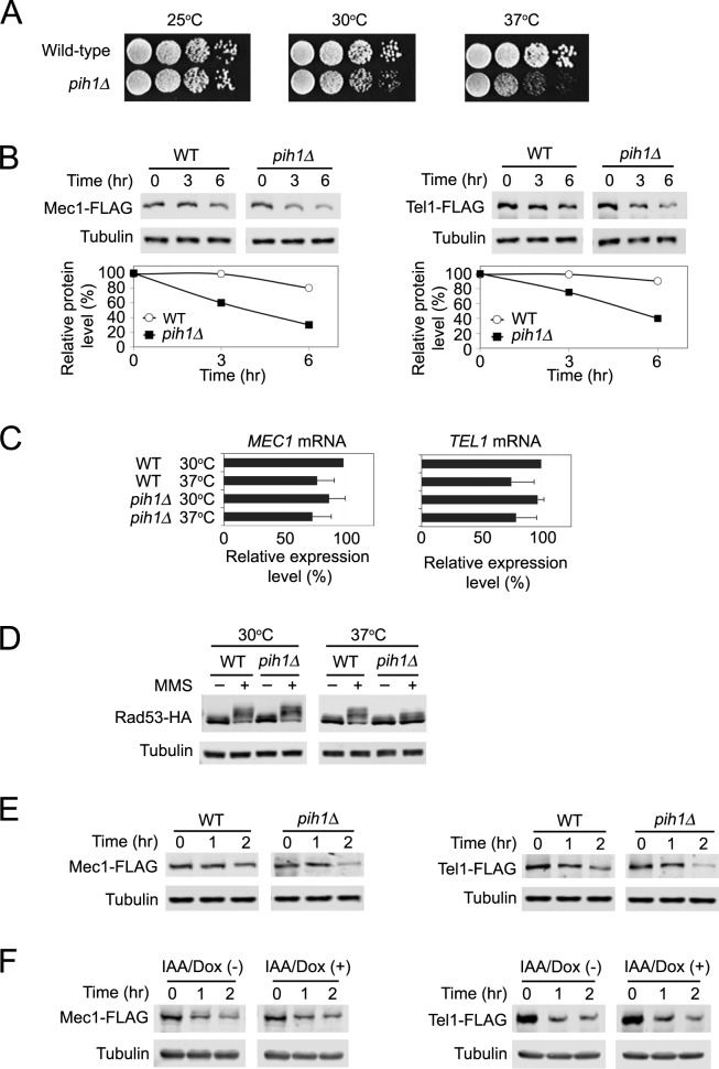
Fig 6. Role of Pih1 in protein stabilization of Mec1 and Tel1 at high temperatures.
(A) Temperature sensitivity of pih1Δ mutants. Ten-fold serial dilutions of cultures were spotted on YEPD medium. Plates were incubated at 25, 30 or 37°C for 2~3 days. (B) Effect of pih1 deletion mutation on endogenous Mec1 and Tel1 protein levels at high temperatures. Wild-type and pih1Δ cells expressing Mec1-FLAG or Tel1-FLAG were grown at 30°C and transferred to 37°C for the indicated times. Cells were subjected to immunoblotting analysis with anti-FLAG or anti-tubulin antibodies. (C) Effect of pih1 deletion mutation on MEC1 and TEL1 mRNA levels at high temperatures. Wild-type and pih1Δ cells were grown at 30°C and then transferred to 37°C for 3 hr. Cells were analyzed as in Fig 1D. (D) Effect of pih1 deletion on Rad53 phosphorylation. Wild-type and pih1Δ cells expressing Rad53-HA were arrested with nocodazole and exposed to MMS at 30°C (left panel). Arrested cells were also transferred to 37°C for 6 hr and exposed to MMS (right panel). Cells were then analyzed by immunobloting with anti-HA or anti-tubulin antibodies. (E) Effect of pih1Δ mutation on pre-synthesized Mec1 and Tel1 protein stability at high temperatures. Wild-type and pih1Δ cells, carrying the GAL-FLAG-MEC1 or the GAL-FLAG-TEL1 plasmid, were initially grown in galactose medium to induce Mec1 and Tel1 expression from the GAL1 promoter at 30°C. Cells were then incubated in 2% glucose to turn off the GAL1 promoter and allow protein maturation of Mec1 and Tel1 at 30°C. After 6 hr incubation with glucose, cultures were treated with cycloheximide and transferred to 42°C (Time point 0 hr). Cells are collected at the indicated time and examined by immunoblotting with anti-FLAG or tubulin antibodies. (F) Effect of Asa1 depletion on pre-synthesized Mec1 and Tel1 protein stability at high temperatures. asa1-aid cells, carrying the GAL-FLAG-MEC1 or the GAL-FLAG-TEL1 plasmid, were initially grown in galactose medium to induce Mec1 and Tel1 expression from the GAL1 promoter at 30°C. Cells were then incubated in 2% glucose to turn off the GAL1 promoter and allow protein maturation of Mec1 and Tel1 at 30°C for 6 hr. Cultures were then treated with Dox/IAA for one hour and subsequently transferred to 42°C or retained at 30°C (at time point 0 hr). Cells are collected at the indicated time and examined by immunoblotting with anti-FLAG or tubulin antibodies. We note that cells were treated as in Figs 1G and 4H before “time point” 0 hr (See S7 and S17 Figs).

