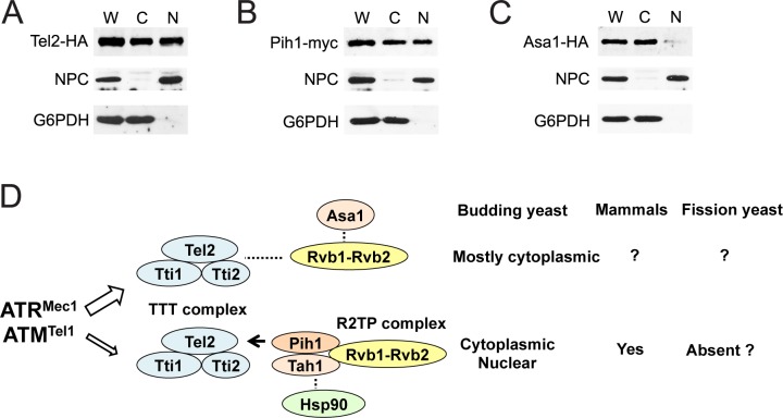
Fig 7. Cellular localization of Asa1 and Pih1.
(A, B, C) Cells expressing Tel2-HA (A), Pih1-myc (B) or Asa1-HA (C) were grown to mid log-phase and spheroplasted. Spheroplasts were homogenized to prepare whole-cell extracts (W) and then separated into the cytoplasmic (C) and nuclear (N) fractions. Samples from each fraction were separated by SDS-PAGE and immunoblotted with anti-HA, anti-myc, anti-Zwf1 (Glucose-6-Phosphate Dehydrogenase; G6PDH) or anti-nuclear pore complex (NPC) antibodies. (D) Two different Tel2 pathways and protein localization. See the text.

