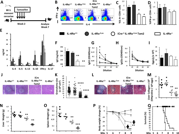
Fig 4. Immunological and histopathological profile of S. mansoni-infected mice after knocking down IL-4Rα 2 weeks post-infection.
A. Experimental design. B. Representative plot of MLN cytokine-producing CD3+CD4+ T cell frequencies after stimulation with PMA/Ionomycin/Monensin cocktail. Summaries of IL-4-producing (C) and IFNγ-producing (D) CD3+CD4+ T cell frequencies from 2 independent experiments conducted with 3–8 mice are shown. E. Cytokine release detected by ELISA in the supernatant of anti-CD3 stimulated MLN cells. F. Total seric IgE in S. mansoni-infected mice. SEA-specific seric IgG1 (G) and IgG2a (H) isotype antibodies. I. Liver Egg burden. J. Formalin-fixed Hematoxylin/Eosin-stained sections of liver tissue from infected animals for morphological analyses (displayed here at 100X). K. Area sizes of egg-surrounding granuloma are computed. L. Formalin-fixed CAB-stained sections of liver tissue from infected animals for collagen detection (displayed here at 100X). M. Hydroxyproline content measured by colorimetry is displayed as a measure of tissue collagen content. Liver (N) and Spleen (O) weights. P. Body weight change over time following S. mansoni infection. Q. Survival curve following S. mansoni infection. Each experiment was conducted at least twice with 5–10 mice per group. Data are expressed as mean ± SD; NS = p > 0.05; * = p < 0.05; ** = p < 0.01; *** =, p < 0.001; **** = p < 0.0001.

