
Figure 1. A CRISPR–Cas9 genetically engineered mouse model for MERS-CoV replication.
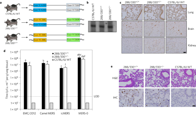
a, C57BL/6J mice were genetically engineered using CRISPR–Cas9 genomic editing to encode 288L and 330R in mDPP4 on one chromosome (heterozygous, 288/330+/−) or on both chromosomes (homozygous, 288/330+/+). b, Northern blot of mDPP4 mRNA expression. c, Immunohistochemistry (IHC) of mDPP4 protein in the lungs, brain and kidneys of individual C57BL/6J wild-type (WT), 288/330+/− and 288/330+/+ mice. d, Viral titres for MERS-CoV at 3 days post-infection from C57BL/6J WT, 288/330+/− and 288/330+/+ (all n = 4) mice infected with 5 × 10⁵ plaque-forming units (p.f.u.) of the indicated viruses. Bar graphs show means + s.d. Student's t-test was used to calculate P < 0.05 for comparisons of MERS-0 with each virus in 288/330+/+ mice (a–c on graph) and 288/330+/− mice (d–f on graph). e, Pathology of 288/330+/+, 288/330+/− and C57BL/6J WT mice at day 3 after infection with MERS-0. Lung tissue sections were stained to examine the pathology by haematoxylin and eosin staining (H&E), or were stained by IHC to detect nucleocapsid protein from MERS-0 infection. IHC and H&E pathology images are representative of at least three samples. Scale bars (c,e), 1 mm.