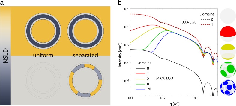
Fig. 2.
Lateral organization of membranes investigated with neutron scattering. a Schematic illustration of selective lipid deuteration and solvent contrast matching to facilitate the detection of membrane domains. b Calculated scattering curves for vesicles with the same average bilayer NSLD, but under different conditions of solvent NSLD and/or lipid lateral organization. Scattering curves were calculated with an analytical form factor for phase-separated vesicles described in Heberle et al. (2015), using 100 expansion orders and model parameters stated in the main text. Figure adapted from Marquardt et al. (2015)

