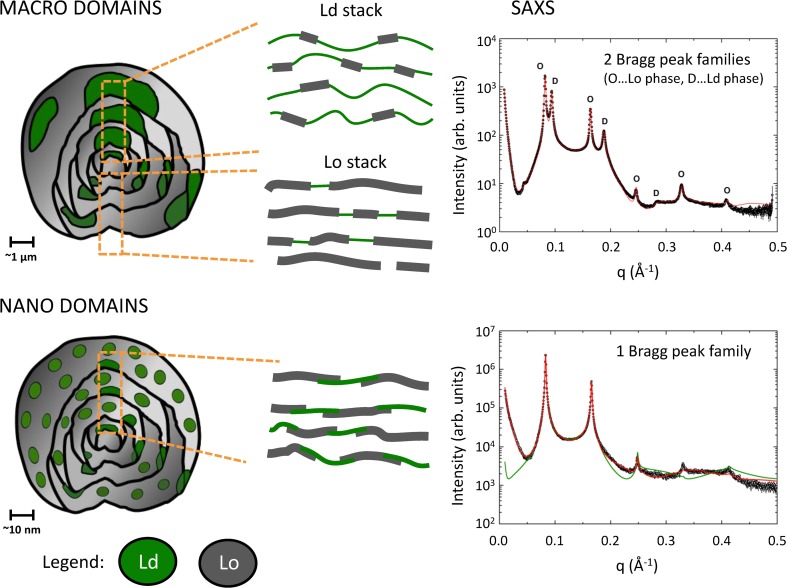
Fig. 6.
Macroscopic and nanoscopic domains in MLVs, in DSPC/DOPC/Chol = 30/46/24 mol% and DSPC/POPC/Chol = 39/39/22 mol%, respectively, at 20 °C. Macroscopic domains show long-range alignment of like domains leading to SAXS pattern with two lamellar lattices. The model fit (red solid line) also considers contributions from positionally uncorrelated Lo domains in Ld stacks and vice versa. SAXS data of nanoscopic domains show only a single lamellar lattice signifying the absence of domain alignment. The underlying model (red solid line) considers partial overlap of Lo and Ld domains. A homogenous (ideally mixed) model of membrane structure was unable to fit experimental data. Data taken from Belička et al. (2017)

