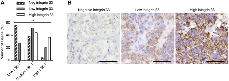
Figure 4.
The LSD1-integrin β3 axis in NSCLC adenocarcinoma. (A) Immunohistostaining of integrin β3 on 182 lung adenocarcinomas was classified into three integrin β3 expression groups (negative, low, and high). Chi-Square tests were used to calculate the statistical significance for linear-by-linear association. p = 0.001. Original data can be found in Supplementary Table S2. (B) Representative images of negative, moderate and strong immunohistochemical stainings of integrin β3. Scale bars, 50 µm.

