Figure 1.
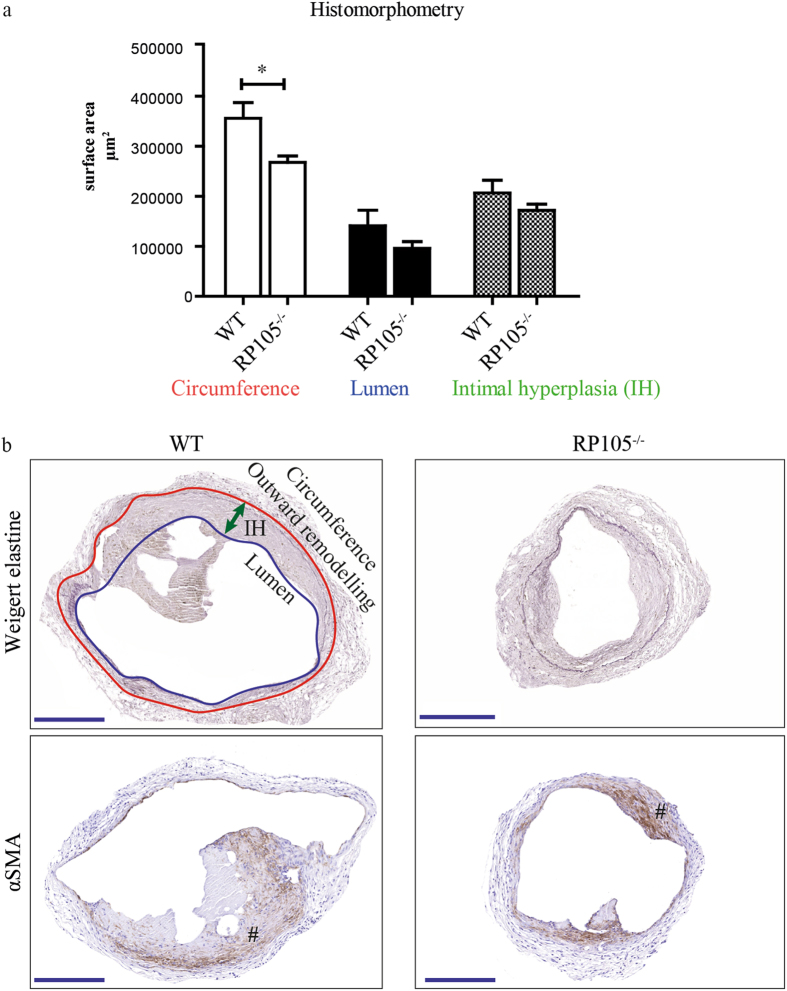
Effect of RP105 deficiency on AVF maturation in vivo. (a) Quantification of morphometric parameters. Decrease in vessel circumference (outward remodeling) in RP105 deficient mice was observed 14 days after AVF creation, compared to WT. Lumen and intimal hyperplasia did not differ between RP105−/− and WT mice. (b) Histological staining of venous outflow tract 14 days after surgery. Weigert elastine staining was used to determine histomorphometrical parameters of the vessel. Circumference (internal elastic lamina area) was used to quantify outward remodeling (red line). Intimal hyperplasia (green arrow) measured as a difference between luminal area (blue line) and vessel circumference. αSMA staining shows area of intimal hyperplasia 14 days after AVF creation. (#) intimal hyperplasia; P < 0.05; n = 11 per group. Bar = 200 μm; 100x magnification.

