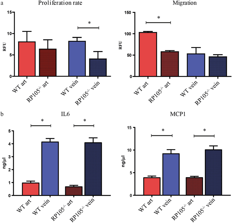
Figure 5.
Functional difference between arterial and venous VSMCs in vitro. Reduction in proliferation rate was limited to VSMCs isolated from RP105−/− veins. Decrease in migration of VSMCs isolated from RP105−/− mice was restricted to arterial cells only. Proliferation rate and migration were measured over 16 h time period. (b) Venous VSMCs isolated from WT and RP105−/− mice produce significantly higher amounts of inflammatory cytokines IL6 and MCP1. Cells were maintain in culture for 14 days. *P < 0.05; n = 3.

