Figure 1.

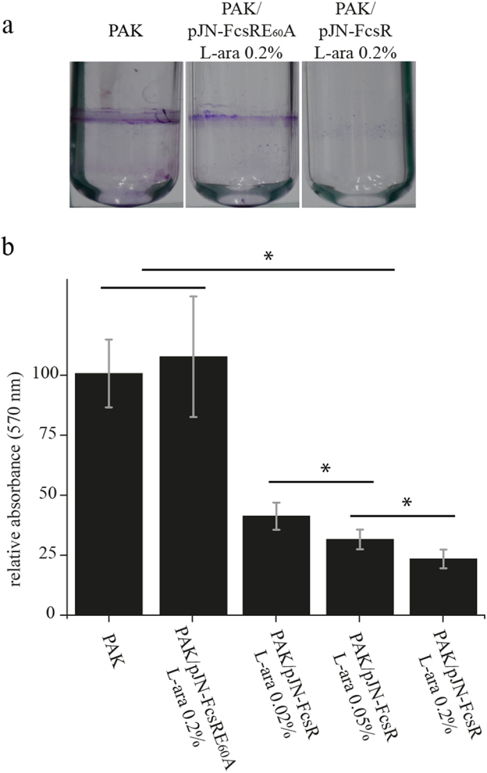
PAK/pJN-FcsR is impaired for biofilm formation. (a) Pellicle formation assays. The figure shows the pellicles associated to the glass tubes stained with crystal violet after removal of the liquid culture. This assay was performed for PAK, PAK/pJN-FcsR and PAK/pJN-FcsRE60A. The concentrations of L-arabinose used as inducer are indicated. (b) Biofilm formation assays. Biofilm formation on solid surface was evaluated using microtiter dish binding assay for PAK, PAK/pJN-FcsR and PAK/pJN-FcsRE60A. L-arabinose concentrations used are indicated. Crystal violet staining retained on the biofilms was measured spectrophotometrically at 570 nm for quantification. *Indicates statistically significant difference determined by ANOVA and Tukey’s post hoc test (p < 0.05).
