Fig. 3.
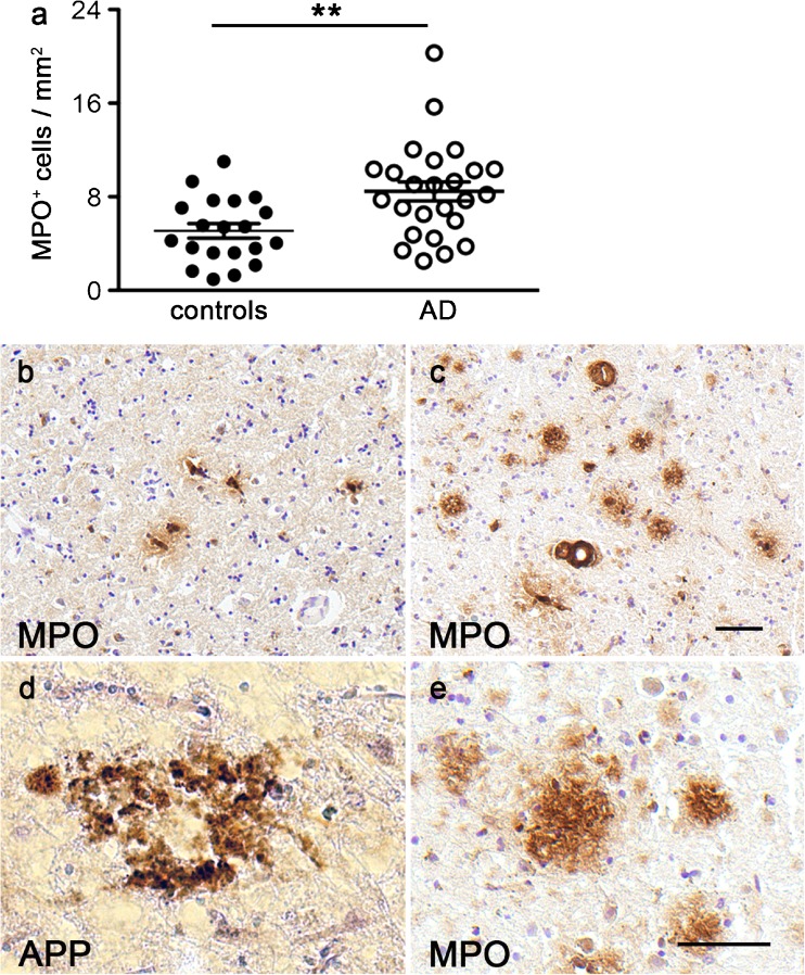
Increased MPO expression in frontal cortex samples from AD patients. The number of MPO-immunoreactive cells was significantly higher in the frontal cortex of AD patients compared with controls (P = 0.003, t = 3.17, df = 63, in a). Examples of MPO-immunoreactive cells are shown in the frontal cortex of a control case (b) and an AD patient (c). Many extracellular protein aggregates show immunostaining for amyloid beta (APP, d) and also for MPO (e) in samples from AD patients. Bars 25 μm (b, c; d, e). ***P < 0.005

