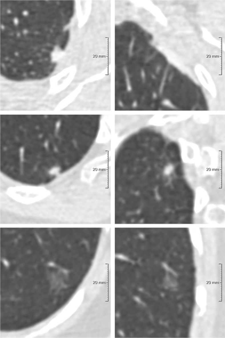
Fig. 2.
Example of one participant in which three different nodules were considered as the risk-dominant lesion. Each row depicts a nodule, displayed in the axial (left) and coronal (right) plane and centered in the images with a field of view of 60 x 60 mm. (A) Solid benign nodule, DmeanAxial 6.1 mm, risk-dominant nodule for the NCCN guidelines with category 5; (B) Solid benign nodule, Dmean3D 8.7 mm, risk-dominant nodule for Lung-RADS with category 4A; (C) Pure ground-glass benign nodule, Dlongest-C 14.3 mm, risk-dominant nodule for the PanCan model with nodule risk index of 0.14%

