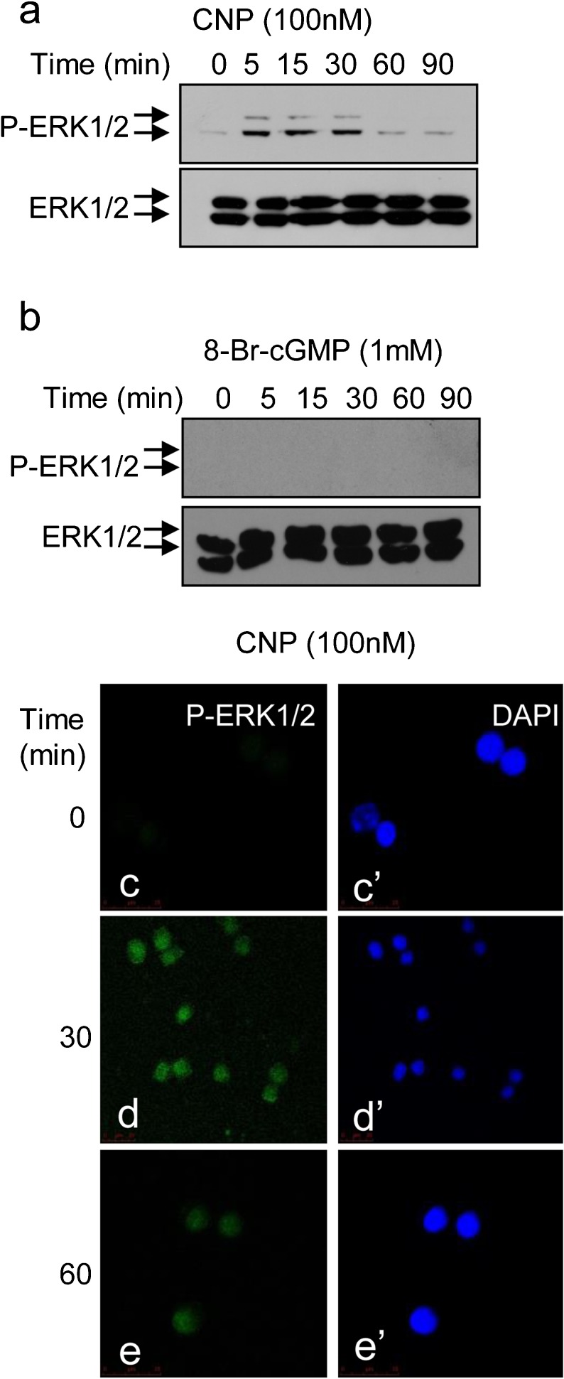
Fig. 2.
Time-course analysis of CNP and dibutryl-cGMP (db-cGMP) effects on ERK1/2 phosphorylation in GH3 cells. a, b GH3 cells were stimulated with (a) CNP (100 nM) or (b) db-cGMP (8-Br-cGMP; 1 mM) for up to 90 min, prior to extraction of total proteins and Western blotting for ERK1/2 phosphorylation. Each autoradiograph is representative of at least three independent experiments. c–e’ GH3 cells were treated for up to 90 min with 100 nM CNP prior to being fixed and stained for phospho-ERK1/2 (Alexa-488, green) or nuclear co-stained (DAPI 4,6-diamidino-2-phenylindole, blue). Immunofluorescence was visualised using confocal microscopy. Images shown are a representative field of vision from at least three independent experiments

