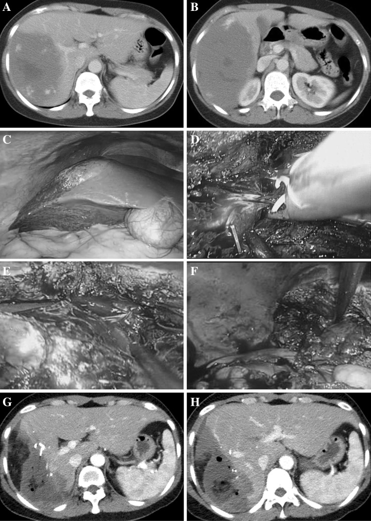
Fig. 2.
Successfully performed enucleation for giant liver hemangiomas with tumor diameter of 21.7 cm in the right posterior segment and reduced excessive loss of normal liver tissue. A, B typical features of hemangioma with contrast-enhanced CT; C tumor exterior characteristics with a laparoscopic horizon; D branches of middle the hepatic vein ligate with Hem-o-lok; E enucleation of hemangiomas along the tumor margin; F the cut surface of the remnant liver; G postoperative contrast-enhanced CT of tumor enucleation without excessive loss of normal liver tissue; H Retained right anterior portal branches without injury

