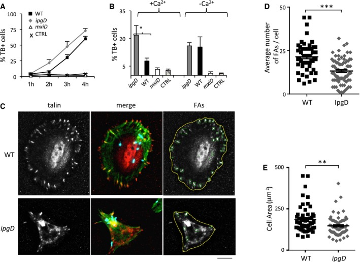
Figure 6. IpgD prevents Ca2+‐dependent early cytotoxicity linked to cell detachment induced by Shigella .

- Cells were challenged with wild‐type Shigella (black squares) or the ipgD mutant (gray diamonds) for the indicated time points, and non‐viable cells were scored by trypan blue staining. Representative experiments performed with triplicate determinations, n > 1,000 cells per point. The results are expressed as the average percentage of trypan blue‐positive cells ± SEM.
- Average percentage of trypan blue‐positive cells ± SEM following bacterial challenge with the indicated strains for 2 h in the absence (−Ca2+) of presence of extracellular Ca2+ (+Ca2+). CTRL: non‐infected cells. N = 3, > 30 cells. Unpaired t‐test, *P = 0.0283.
- Talin‐GFP‐transfected cells were challenged with the indicated Shigella strains for 1 h and processed for immunofluorescence staining. Talin: representative projection of deconvolved epifluorescence microscopy planes showing cells challenged with the indicated Shigella strains. Merge: actin: red, talin: green; bacteria: blue. FAs: detection of FAs performed using an automated algorithm (Materials and Methods). Scale bar, 5 μm.
- The average number of FAs/cell ± SEM was determined for cells infected with the indicated bacteria as in (C). N = 3, > 30 cells. Unpaired t‐test, ***P ≤ 0.001
- The average cell area ± SEM was determined for cells infected with the indicated bacteria as in (C). N = 3, > 30 cells. Wilcoxon test, **P = 0.0031.
