
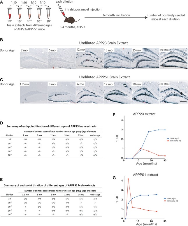
Figure 2. Seeding activity of brain extracts from APP23 and APPPS1 tg mice increases with donor age and plateaus in late stages.
-
A–CBrain extracts from APP23 (B) and APPPS1 (C) mice at different ages were injected into the hippocampus of young, pre‐depositing 3‐ to 4‐month‐old male APP23 host mice. Brains were immunohistochemically analyzed for Aβ deposition 6 months later. Aβ immunostaining combined with Congo red staining is shown. Note the more diffuse and filamentous Aβ deposition induced with the APP23 extracts in contrast to the punctate and compact Aβ deposition induced with the APPPS1 extracts. Scale bars: 200 μm.
-
D, ENumber of mice with induced Aβ deposition at each dilution from the different age groups (n = mainly 3–6/group) of APP23 (D) and APPPS1 (E) brain extracts.
-
F, GSD50 of APP23 (F) and APPPS1 (G) brain extracts (blue line; Reed‐Muench method, see Table 1 for statistical analysis). SD50 (half‐maximal seeding dose) was defined as the log10 of the brain extract dilution at which 50% of the host mice showed induced Aβ deposition (see Materials and Methods). The specific seeding activity (SD50/total Aβ; red line) for each extract indicates a peak at early ages in brain extracts from both mouse lines (see Table 2).