
Figure EV4. Analysis of miRNA and IL‐7R expression in the absence of IL‐7 and pre‐BCR signaling as well as in a miR‐15 loss‐of‐function/gain‐of‐function setting.
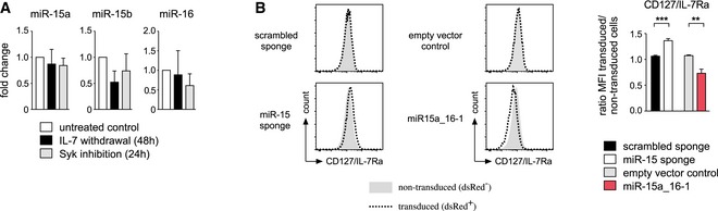
- Increased miRNA activity in differentiating pre‐B cells is not accompanied by increased miRNA expression. Total RNA isolated from wk3 cells cultured without IL‐7 for 48 h, with the Syk inhibitor R406 (2 μM) for 24 h or left untreated was converted into cDNA, and expression of miR‐15a, miR‐15b, miR‐16, and sno‐202 as a reference was determined by quantitative PCR. Bars display the fold change, normalized to the untreated control sample (set as 1). Values are expressed as mean ± SD and represent four independent experiments.
- IL‐7R surface expression is regulated by the activity of the miR‐15 family. Pre‐B cells expressing the miR‐15 sponge or miR‐15a‐16‐1 as indicated were analyzed for surface IL‐7R as measured by CD127/IL‐7Ra expression. Histograms display a representative experiment, comparing non‐transduced (gray graph) and transduced cells (dotted line) within the same sample. The bar graph shows the corresponding means ± SD of four independent experiments, in each case compared by a paired t‐test; ***P < 0.001, **P < 0.01.