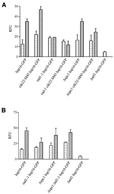
Figure 3.
Hsp16–GFP expression is activated following HU or CPT treatment. Analysis of hsp16–GFP levels in cdc22-M45 mutant. (A) Cells were cultured at 25°C (lightly shaded bars) in YEA to mid-exponential phase and then hydroxyurea (heavily shaded bars) was added at a final concentration of 11 mM to one-half of the culture and incubated for 4 h. (B) Cells were cultured at 25°C (lightly shaded bars) in YEA to mid-exponential and then camptothecin (heavily shaded bars) was added at a final concentration of 40 µM to one-half of the culture and incubated for 2 h. Hsp16–GFP levels were quantitated by fluorimetry.

