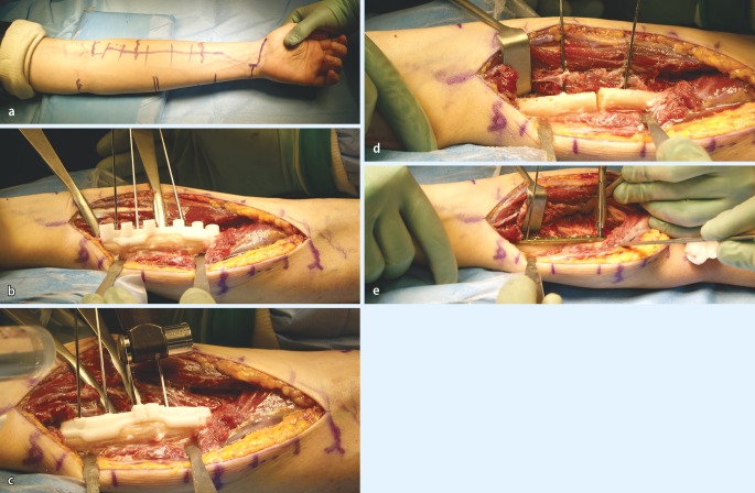
Fig. 7.
Clinical intraoperative images of the patient, whose planning is depicted in Fig. 4. a Marking of the palmar Henry approach to the radius. b After visualization of the profound branch of the radial nerve the rapid prototyping drill template is placed on the apex of the deformity. c After exchanging of the drill template to the cutting template, the osteotomy is performed. d The osteotomy is complete. e Sound osteosynthesis with a 6 hole modern osteosynthesis plate, using the predrilled holes to restore anatomy

