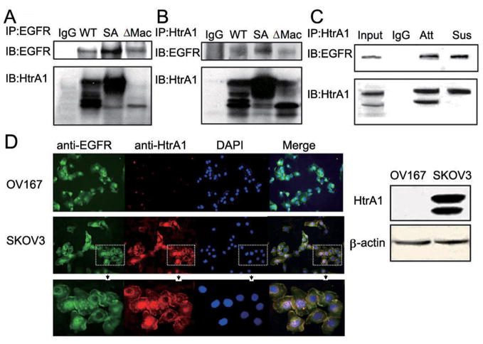
Figure 5.
HtrA1 interacts with EGFR. A-B, exogenous HtrA1 and EGFR associate with each other. Plasmids encoding WT HtrA1, SA mutant HtrA1 or ΔMac25 HtrA1 were co-transfected with EGFR separately into HEK293T cells. After 24 h, immunoprecipitation was performed with mouse anti-EGFR antibody (A) or rabbit anti-HtrA1 antibody (B). Normal IgG was used as the negative control. C, endogenous HtrA1 associates with endogenous EGFR. SKOV3 cells were cultured under attachment condition (Att) or suspension condition (Sus) for 8 h, followed by immunoprecipitation with rabbit anti-HtrA1 antibody. D, immunolocalization of HtrA1 and EGFR. SKOV3 cells were fixed with 4% PFA and stained for the expression of EGFR (green) and HtrA1 (red). Nuclei were counterstained with DAPI (blue). OV167 cells which do not express HtrA1 served as negative controls. HtrA1 co-localizes with EGFR on the cell membrane in SKOV3 cells (see merged images). The bottom panel shows magnification of regions in dashed square from the middle panel. The right panel shows expression levels of HtrA1 in OV167 and SKOV3 cells.

