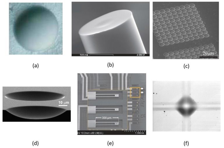
Figure 5.
Microscope images of (a) a dimple with diameter of 200 μm formed by a bubble-trapping method [76]; (b) a laser machined fiber end facet [12]; (c) array of concave features made by FIB milling [11]; (d) two silicon micro-mirrors fabricated by dry etching [78]; (e) array of cantilever based micro-mirrors [79], (f) and a microscope image of a waveguide connected buckled-dome microcavity. The dome diameter is 100 μm [80].

