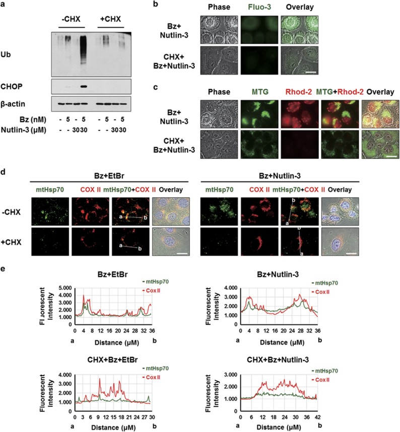
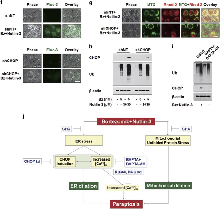
Figure 8.
Upregulation of CHOP and the increase in Ca2+ levels precede the ER dilation induced by bortezomib/nutlin-3. (a) MDA-MB 435S cells pretreated with or without 2 μM CHX and further treated with 5 nM bortezomib plus 30 μM nutlin-3 for 16 h. Cell extracts were prepared for the western blotting of ubiquitin and CHOP. β-actin was used as a loading control in western blots. (b, c) MDA-MB 435S cells were pre-treated with or without 2 μM CHX for 30 min further treated with 5 nM bortezomib and 30 μM nutlin-3 for 16 h. (b) Treated cells were stained with 1 μM Fluo-3 and observed under the phase-contrast and fluorescence microscope. Scale bar, 20 μm. (c) Treated cells were stained with 1 μM Rhod-2 and 100 nM MTG and observed under the phase-contrast and fluorescence microscope. Scale bar, 20 μm. (d) MDA-MB 435S cells pretreated with or without 2 μM CHX followed by treatment with 5 μg/ml EtBr for 24 h. After culture media was changed with the fresh ones, cells were further treated with 5 nM bortezomib for 12 h. Treated cells were fixed, and subjected for immunocytochemistry of COX II and mtHsp70. Scale bars, 20 μm. (e) Plots of the intensity profile of a set of pixels distributed on a lline drawn across the indicated mitochondria (as shown in mtHsp70+COX II panels) at differnet emission wavelengths corresponding to the signals of mtHsp70 (green) and COX II (red). (f–h) MDA-MB 435S cells were infected with the lentivirus containing non-targeting (NT) shRNA or a CHOP-targeting shRNA (CHOP shRNA) for 24 h. Infected cells were treated with 5 nM bortezomib and 30 μM nutlin-3 for 16 h. (f) Treated cells were stained with 1 μM Fluo-3 and observed under the phase-contrast and fluorescence microscope. Scale bars, 20 μm. (g) Treated cells were stained with 1 μM Rhod-2 and 100 nM MTG and observed under the phase-contrast and fluorescence microscope. Scale bars, 20 μm. (h) Cell extracts were prepared for western blotting of CHOP and ubiquitin. β-actin was used as a loading control in western blots. (i) MDA-MB 435S cells pretreated with 10 μM BAPTA and 10 μM BAPTA-AM were further treated with 5 nM bortezomib plus 30 μM nutlin-3 for 8 h and then western blotting of the indicated proteins was performed. β-actin was used as a loading control in western blots. (j) Hypothetical model of the underlying mechanism of paraptosis induced by bortezomib/nutlin-3 in p53-defective cancer cells. Combination of bortezomib and nutlin-3 triggers ER stress induction, CHOP upregulation, and the increase in cytosolic Ca2+ levels, leading to ER dilation. In parallel to ER dilation, mitochondria are also dilated possibly via the mechanism related to mitochondrial unfolded protein stress, contributing to the paraptotic cell death induced by bortezomib/nutlin-3. Mitochondrial Ca2+ overload, which is preceded by increased cytosolic Ca2+, may also play a role in the cytotoxicity of bortezomib/nutlin-3.


