Figure 2.
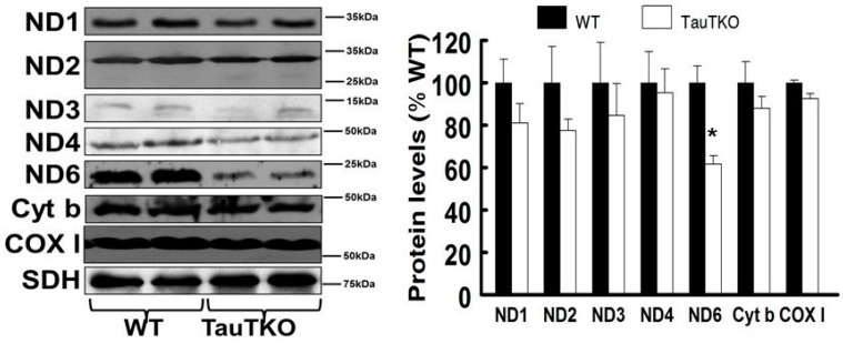
ND6 is reduced in TauTKO hearts. The mitochondrial fraction was isolated from homogenized hearts and then subjected to western blot analyses. The left panel shows representative gels for ND1, ND2, ND3, ND4, ND6, Cyt b (cytochrome b) and COX I (cytochrome c oxidase I), with succinate dehydrogenase (SDH) serving as the loading control. The right panel shows the means ± SEM of the mitochondrial protein/SDH ratio of 6–9 different hearts. Values are expressed relative to wild-type (WT), where WT is fixed at 100%. * p < 0.05.

