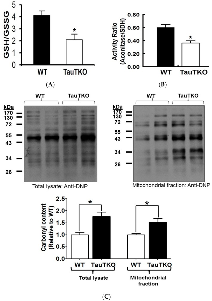
Figure 4.
Taurine depletion causes oxidative stress. (A) Reduced (GSH) and oxidized (GSSG) glutathione content were determined and the data expressed as the glutathione redox state (GSH/GSSG), with values shown representing means ± SEM of 6–8 hearts. * p < 0.05; (B) Aconitase activity of WT and TauTKO mitochondria were assayed and normalized relative to SD activity. Values represent means ± SEM of 4–6 hearts. * p < 0.05; (C) Following preparation of total heart lysates and the mitochondrial fraction, proteins were derivatized with 2,4-dinitrophenylhydrazine and then subjected to western blot analysis of carbonylated proteins. The top panels show representative gels of carbonylated proteins of total lysate and the mitochondrial fraction. Values shown in the bottom panel represent means ± SEM for relative cellular and mitochondrial carbonylated protein content from 4–5 hearts. Values are expressed relative to WT, where WT is fixed at 1.0. * p < 0.05.

