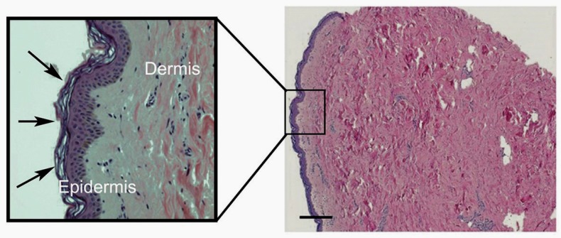
Figure 1.
Micrograph of human breast skin sample, showing the full depth of the dermis (pink staining) in comparison to the thin layer of epidermis (purple staining). The scale bar indicates 200 µm. A zoomed-in image is shown within the box. The stratum corneum, the outermost layer of the epidermis, is indicated by the arrows, with its characteristic basket-weave structure. The collagen bundles in the dermis are very clear, as are the scattered purple-stained fibroblasts that generate this structure.

