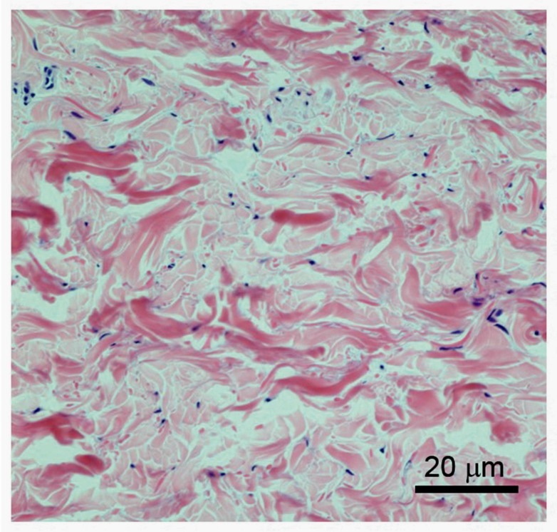
Figure 3.
Structure of the dermis. Higher magnification of H&E-stained dermis, showing the irregular nature of the bundled collagen fibres (pink stained) and sparse presence of the fibroblasts (blue nuclear staining). Vitamin C present in the fibroblasts supports the synthesis of the collagen fibres.

