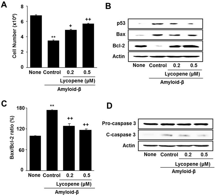
Figure 2.
Effect of lycopene on cell viability and apoptotic indices in amyloid-β-stimulated cells. The cells were pretreated with lycopene for 1 h and then stimulated with amyloid-β (20 μM) for another 24 h. (A) Viable cell numbers was determined by the Trypan Blue exclusion test; (B) Levels of p53, Bax, Bcl-2, caspase-3, and actin were determined by western blot analysis; (C) The ratio of Bax/Bcl-2 was determined by protein band densities of Bax and Bcl-2; (D) Levels of pro- and cleaved-caspase-3 were assessed by western blot analysis. Actin was used as a loading control. The value of None (without any stimulation or treatment) was set as 100%. Data are expressed as the mean ± S.E. of three independent experiments. ** p < 0.01 vs. None (without any stimulation or treatment); + p < 0.05, ++ p < 0.01 vs. Control (with amyloid-β stimulation alone). c-caspase-3, cleaved caspase-3.

