
Figure 5.
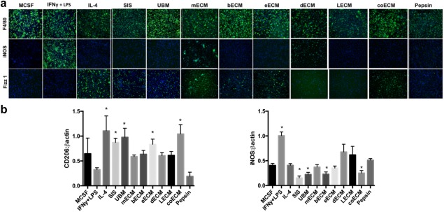
Scaffolds derived from extracellular matrix components of specific tissues polarize macrophage function. (a) Macrophages (stained with F4/80) and treated for 18 hr with control cytokines (MCSF, IFN‐γ + LPS, IL‐4) or solubilized extracellular matrix scaffolds cause differential expression of markers for the pro‐inflammatory M1 macrophage (iNOS) and the wound‐healing M2 macrophage (Fizz1) phenotypes. Solubilized scaffolds were produced via pepsin incubation of small intestine submucosa (SIS), urinary bladder (UBM), skeletal muscle (mECM), brain (bECM), esophagus (eECM), skin (dECM), liver (LECM), or colon (coECM) extracellular matrix components. MCSF was used as a negative control for macrophages while pepsin was a control for ECM solubilization, IFN‐γ + LPS was a positive control for M1 macrophages, and IL‐4 was a positive control for M2 macrophages. (b) Treatment with matrices also induces varied levels of M2 associated (CD206) and M1 associated (iNOS) proteins in macrophage lysates. Adapted with permission from Dziki JL, Wang DS, Pineda C, Sicari BM, Rausch T, Badylak SF. Solubilized extracellular matrix bioscaffolds derived from diverse source tissues differentially influence macrophage phenotype. J Biomed Mater Res. 2017;105(1):138‐147