
Figure 7.
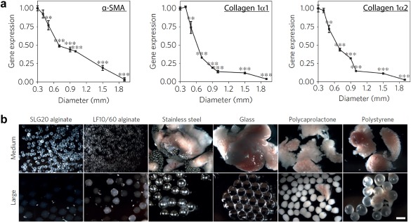
Implanted materials with distinct sizes and compositions alter fibrotic capsule formation. (a) Following implantation of alginate spheres of different sizes into the peritoneum of mice, gene expression profiles of the pro‐fibrotic markers α‐SMA (left), collagen 1α1 (center), and collagen 1α2 (right) revealed larger particles reduced fibrotic build‐up. (b) Images revealing the level of fibrosis for particles with diameters of 0.5 mm (medium) or 1.5–2 mm (large) prepared from alginate, stainless steel, glass, polycaprolactone, or polystyrene. For all materials, large particles were associated with reduced fibrosis 14 days after implantation. (Scale bar, 2 mm). Adapted with permission from Macmillan Publisher Ltd: Nature Materials from Veiseh O, Doloff JC, Ma M, et al. Size‐ and shape‐dependent foreign body immune response to materials implanted in rodents and non‐human primates. Nat Mater. 2015;14(6):643‐651, copyright 2015