. 2017 Jun 25;10(2):107–114. doi: 10.3400/avd.ra.17-00038

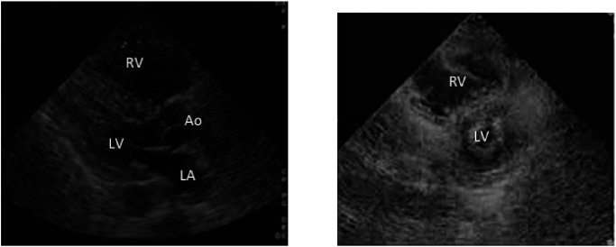
Fig. 2 Right ventricular load in massive pulmonary embolism seen on a trans-thoracic echocardiogram. Left, long-axis view of the heart in a patient with massive pulmonary embolism. Right, short-axis view of the heart in the same patient. Note the enlarged right ventricle and small left ventricle. RV: right ventricle; LV: left ventricle; Ao: ascending aorta; LA: left atrium.

Fig. 2 Right ventricular load in massive pulmonary embolism seen on a trans-thoracic echocardiogram. Left, long-axis view of the heart in a patient with massive pulmonary embolism. Right, short-axis view of the heart in the same patient. Note the enlarged right ventricle and small left ventricle. RV: right ventricle; LV: left ventricle; Ao: ascending aorta; LA: left atrium