. 2017 Mar 31;10(1):44–47. doi: 10.3400/avd.cr.16-00119

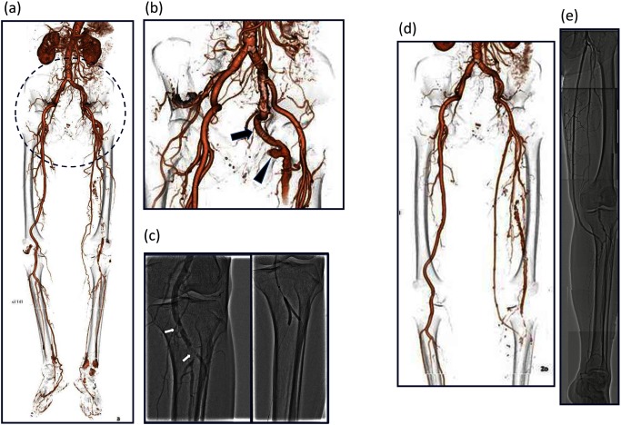
Fig. 1 (a) CT showing a fusiform aneurysmal dilation and occluded PSA. The popliteal artery was fed by collateral circulation. (b) PSA (arrow) and fusiform aneurysmal dilation of PSA (arrowhead). (c) Filling defects of the popliteal artery were detected below the knee. Endovascular treatment of distal embolization from a PSA aneurysm had been performed three times. (d) Postoperative computed tomography showing a patent bypass graft. (e) Postoperative angiography showing a patent bypass graft.

Fig. 1 (a) CT showing a fusiform aneurysmal dilation and occluded PSA. The popliteal artery was fed by collateral circulation. (b) PSA (arrow) and fusiform aneurysmal dilation of PSA (arrowhead). (c) Filling defects of the popliteal artery were detected below the knee. Endovascular treatment of distal embolization from a PSA aneurysm had been performed three times. (d) Postoperative computed tomography showing a patent bypass graft. (e) Postoperative angiography showing a patent bypass graft.