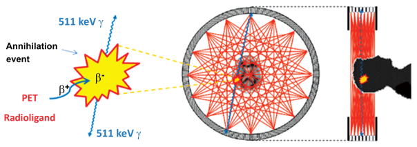
Fig. (1).
Physical basis of PET brain imaging. A PET radioligand that has entered brain after intravenous administration emits a positron. This positron loses kinetic energy over a short distance of a few millimeters before combining with an electron to form positronium, which then annihilates to produce two 511 keV γ-rays that are emitted in almost exactly opposite directions. The PET camera has several adjacent rings of sensitive γ-ray detectors that record all such coincident arrivals of paired γ-rays. Lines between pairs of simultaneously activated detectors cover the positions of positron annihilation events from which PET scans can be reconstructed to measure the distribution of radioactivity in three dimensions. Rapid sequences of scans are recorded to provide regional kinetic information over a few half-lives of the radioligand label (i.e.; 11C or 18F). The detection of coincident γ-ray arrivals amounts to ‘electronic collimation’, and dispenses with the need for ‘physical collimation’ which would otherwise diminish detection sensitivity as in SPECT.

