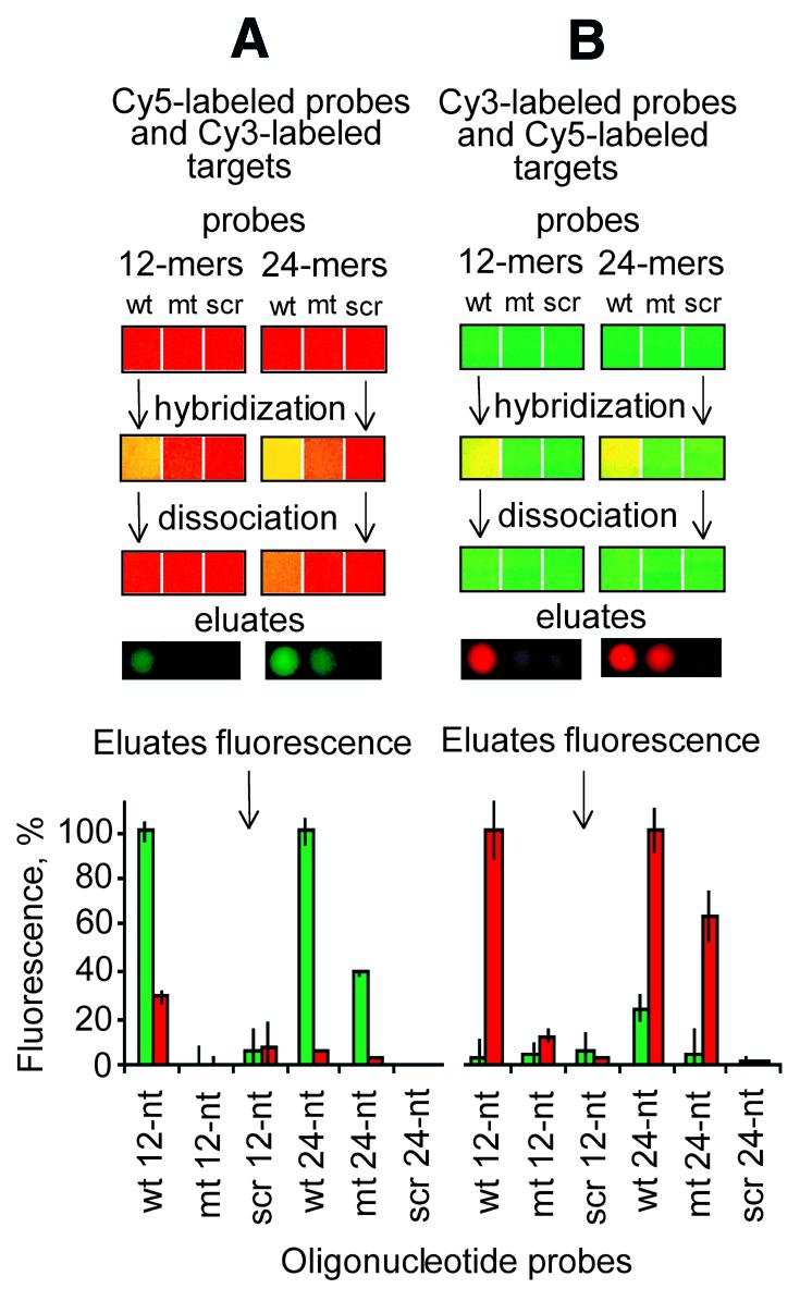
Figure 5.

Analysis of the washing eluate. (A) Patches of aminosilanized surface (3 mm2 each) were saturated with Cy5-labeled probes (red), followed by rinsing to remove excess probe as described in Materials and Methods. Cy3-labeled targets (green) were hybridized to these patches, rinsed to remove the unbound targets and washed in 2 µl of the washing buffer for 15 min (see Materials and Methods). A 0.2 µl aliquot was aspirated from the resulting washing buffer and spotted on a clean slide, which was subsequently imaged with Cy3 and Cy5 filter sets on an ArrayWorx Imager (Applied Precision). (B) The reverse experiments are also shown, where the targets and probes had been reversed. The bar graphs represent the normalized means and the standard deviations of the mean from four such 0.2 µl eluates.
