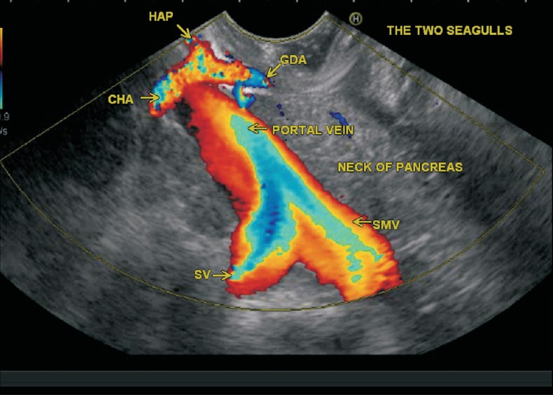
Figure 7.

This image shows two vascular seagulls from duodenal bulb. The upper part of the image showing the division of the CHA into the GDA and HAP gives an appearance of smaller vascular seagull. A larger vascular seagull is also seen due to confluence of SV and SMV
