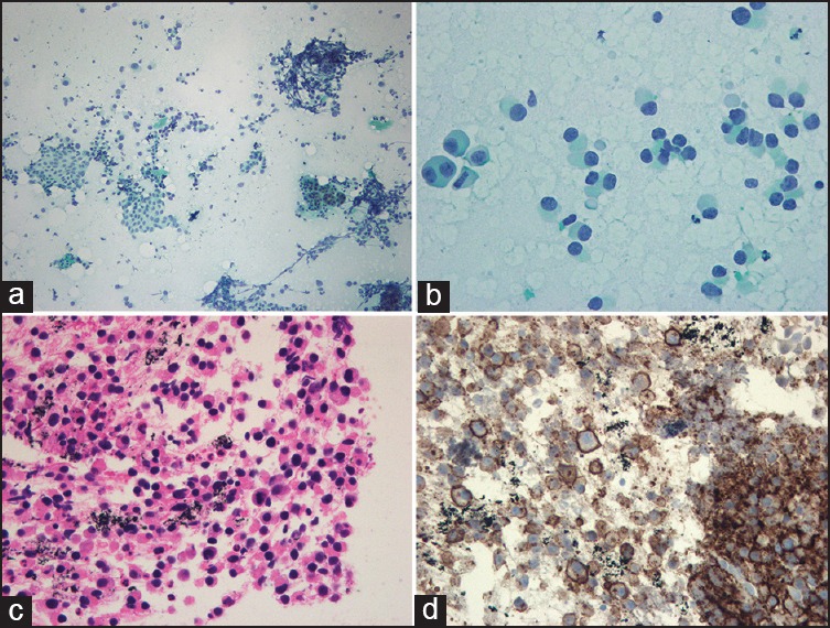
Figure 2.

Pathological findings (a) FNA cytology smear showed a moderately cellular tumor cell population along with normal pancreatic acinar cells and ductal cells (Papanicolaou stain, 100×) (b) At higher magnification, tumor cells showed plasmacytoid appearance with eccentric nuclei and abundant deep basophilic cytoplasm (Papanicolaou stain, 400×) (c) The biopsy specimen demonstrates a sheet of plasmacytoid cells with mild anisonucleosis and mitotic activity (hematoxylin and eosin, 200×) (d) The tumor cells are diffusely positive for plasma cell marker (CD138) (200×)
