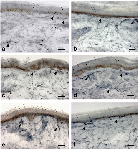
Fig. 2.

Skin biopsy samples at proximal thigh (a-c-e) and distal leg (b-d-f) from a GD1 patients with dying-back skin denervation (a-b), with non lenght-dependent pattern (c-d), and in healthy subject (e-f). Arrows indicate intraepidermal nerve fibers, arrowheads indicate dermal nerve bundles. The density of intraepidermal nerve fibers is reduced in A-B-C. Bright-field immunohistochemistry in 50 μm sections stained with polyclonal rabbit anti- protein-gene-product 9.5 antibody. Bar = 60 μm
