
Fig. 4.
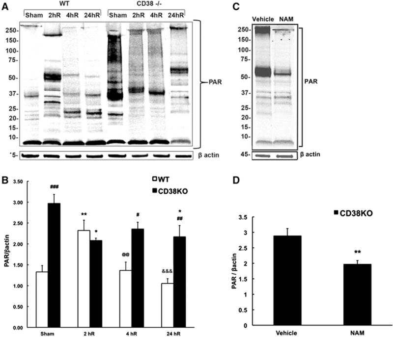
CD38 animals show dramatically higher poly-ADP-ribosylated (PAR) proteins levels when compared to WT animals. a, b There is a significant increase in PAR levels at 2 h (2hR) recovery time in WT mice that then decreased back to sham levels at 4 and 24 h of recovery (4hR, 24hR). The sham CD38KO animals show dramatic increase in PAR levels when compared to sham WT mice. Surprisingly, in CD38KO animals the poly-ADP-ribosylation was reduced following ischemia. b Quantification of western blot images. β actin was used as the loading control. *p < 0.05, **p < 0.01; when compared to corresponding sham, #p < 0.05, ##p < 0.01, ###p < 0.001 when compared to corresponding WT, @@p < 0.01 when compared to 2hR WT, &&&p < 0.001 when compared to 2hR WT, ANOVA followed by Student–Newman–Keuls test, n = 6. c, d Effect of PARP1 inhibitor nicotinamide (Nam) on hippocampal PAR levels. c Immunoblots show marked reduction of band intensities in samples from Nam treated animals. d Nam reduced the hippocampal PAR levels by about 30 %. **p < 0.01 when compared to vehicle, student t test, n = 8