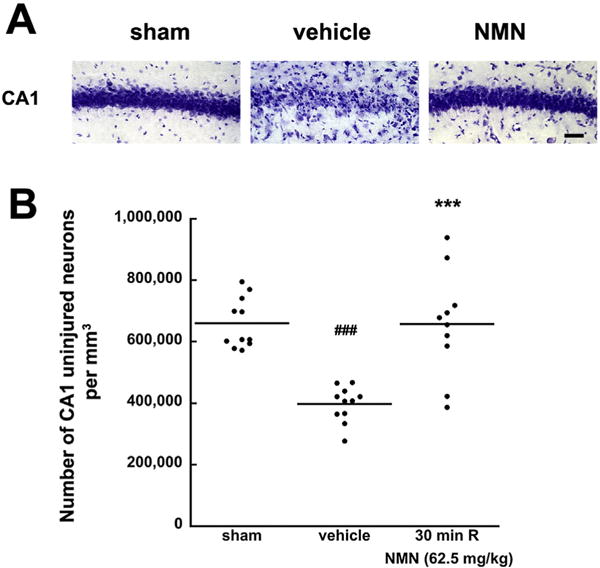
Fig. 2.

Effect of delayed NMN treatment on CA1 neuronal cell death induced by global cerebral ischemia. Cresyl violet staining of the post-ischemic hippocampal CA1 sector following 6 days of recovery (A). In sham operated animals all the CA1 neurons appeared normal. Animals treated with vehicle and NMN (62.5 mg/kg) at 30 min after start of reperfusion. There is a major loss of CA1 pyramidal neurons in vehicle treated animals, while NMN treated animals show normal neuronal morphology. Scale bar represents 50 μm. (B) Shows that there is about 50% cell death at 6 days of recovery in delayed vehicle treated animals. NMN administration at 30 min of recovery significantly improves the cells survival. The horizontal lines represent the mean value. ***p < 0.001 when compared to the vehicle treated group (n = 10), ###p < 0.001 cell loss when compared to sham (n = 10), ANOVA followed by Tukey HSD test.
