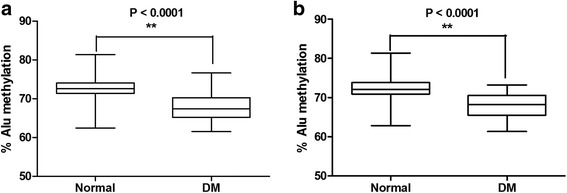
Fig. 4.

The percentage of Alu methylation in age-matched pairs between normal samples and DM samples in both the FBS (a) and HbA1C groups (b). The values from 30 independent age-matched pairs are shown as box plots, with the boxes representing interquartile ranges (25th to 75th percentile) and median lines representing the 50th percentile. The whiskers represent minimum and maximum values. *P < 0.05, **P < 0.001 (Mann-Whitney test)
