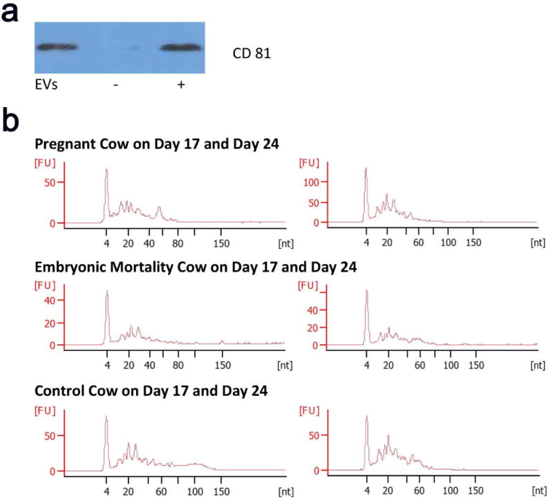
Figure 2.

Evaluation of isolated extracellular vesicles and associated RNA. (a) Representative Western blot showing the presence of CD81, a well characterized marker, in serum-derived extracellular vesicles (EVs) from a cow assigned to the pregnant group, compared to negative (−) and positive (+) controls. All extracellular vesicle samples isolated from the pregnant, embryonic-mortality, and control groups possessed CD81. (b) Agilent electropherogram profiles of small RNAs extracted from circulating extracellular vesicles. The base pair length [nt] (x-axis) is plotted against the fluorescence units [FU] (y-axis). Note the clustering of RNA species below 60 bp in all samples.
