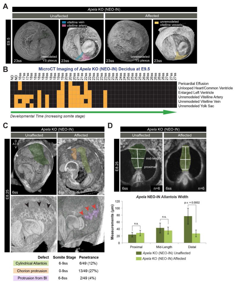
FIGURE 5. Vitelline vessel remodeling defects and extraembryonic anomalies in affected Apela KO embryos.
(A) 3D reconstructions of microCT images at E9.5. Vitelline vessels are pseudocolored: remodeled vitelline vein (blue) and vitelline artery (pink), unremodeled vitelline vessels (yellow). (B) Summary matrix of microCT results at E9.5 from 5 litters (n=46 KO NEO-IN decidua, 3 reabsorbed embryos). X-axis lists individuals by somite stage (ss; ND, not determined). Y-axis lists normal (black boxes) and abnormal (orange boxes) classification of phenotypes. (C) 3D reconstructions of microCT at E8.25 with pseudocolored structures: normal allantois (dark green), cylindrical allantois (light green), ectopic chorion protrusion (orange), normal blood island (BI; black arrowheads), abnormal BI protrusions (purple, with red arrowheads). (D) Posterior views of microCT images used to measure allantois width at E8.25. Data are represented as mean ± SD.

