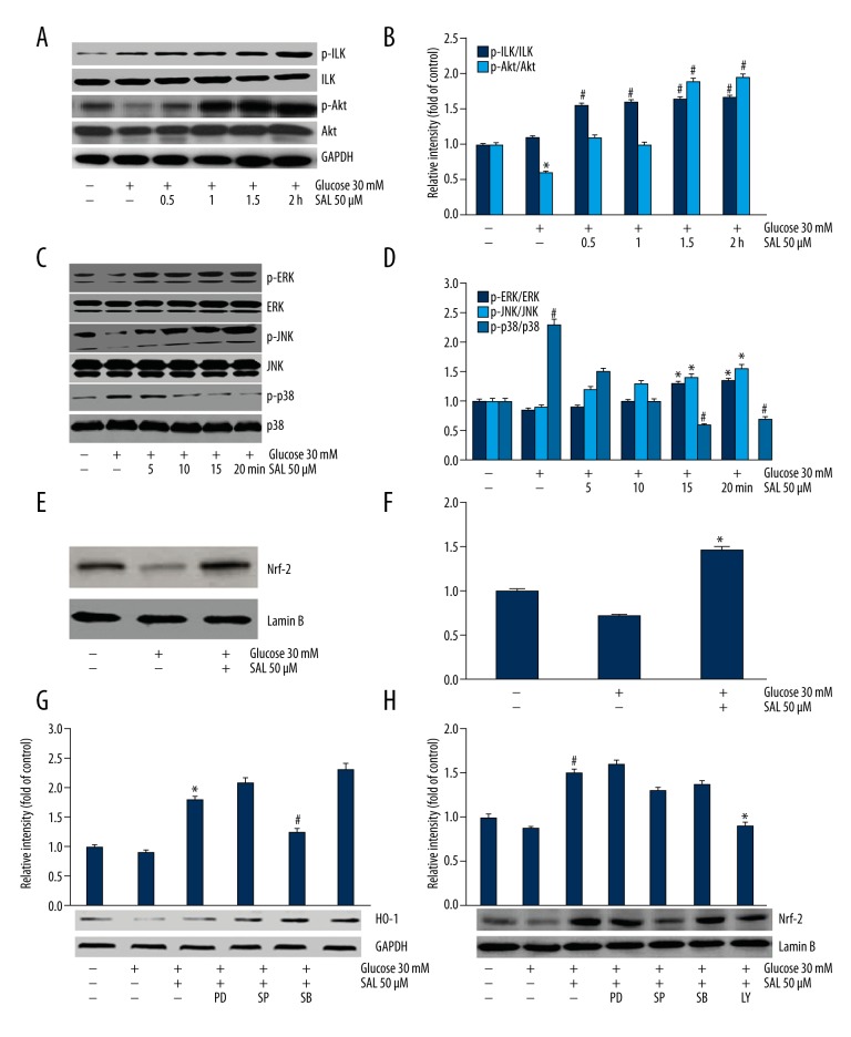
Figure 4.
The involvement of p38, Akt/ILK, JNK and ERK signaling pathways, and Nrf2 nucleus localization was investigated. (A, B) The expression of p-Akt and p-ILK was evaluated using Western blot analysis after culturing in salidroside (50 μM) for 0, 0.5, 1, 1.5, and 2 h. The data were analyzed by ANOVA. * P<0.05 compared with control. # P<0.05 compared with high glucose group. (C, D) The expression of phosphorylated MAPK family members was assessed after culturing in salidroside (50 μM) for 0, 5, 10, 15, and 20 min. The data were analyzed by ANOVA. # P<0.05 compared with control. * P<0.05 compared with high glucose group. (E, F) Nrf-2 nuclear localization was evaluated after treatment with salidroside (50 μM). The data were analyzed by independent-samples t test. * P<0.05 compared with high glucose group. (G) HO-1 expression was assessed after podocytes were treated with ERK1/2 inhibitor PD98059 (25 uM, 0.5 h), JNK inhibitor SP600125 (20 uM, 0.5 h), and p38 inhibitor SB203580 (10 uM, 1 h). The data were analyzed by independent-samples t test. * Compared with high glucose group, # compared with high glucose+salidroside group P<0.05. (H) Nrf2 localization was assessed. * Compared with high glucose+salidroside group. # Compared with high glucose group P<0.05. PD (ERK1/2 inhibitor PD98059), SP (JNK inhibitor SP600125), SB (p38 inhibitor SB203580), LY (ILK inhibitor LY294002). At least 3 independent experiments with 3 replicates per experiment were conducted.

