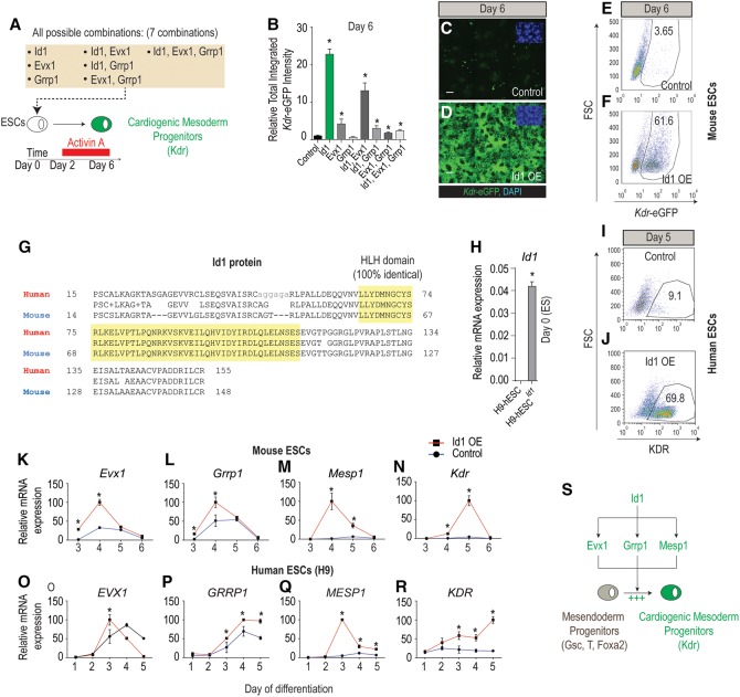
Figure 2.
Id1 is sufficient to direct Kdr+ mesoderm formation in mESCs and hESCs. (A) Schematic of the strategy to evaluate the sufficiency (gain of function) of any of three candidates alone or in combination to promote mesoderm differentiation. (B) Kdr-eGFP fluorescence measurement at day 6 of differentiation in mESCs overexpressing all possible combinations of the three candidates plotted relative to uninfected control levels. (C,D) Representative images of Kdr-eGFP for Id1-overexpressing versus control mESCs illustrating the results presented in B. Bar, 50 µm. (E,F) Flow cytometry analysis reveals that 61.6% of Id1-overexpressing mESCs differentiate into Kdr-eGFP+ mesoderm as compared with 3.65% for control cells at day 6. (G) Alignment and comparison of the mouse (NP_034625.1) Id1 HLH domain and the human (NP_851998.1) Id1 HLH domain using the Protein Blast tool (https://blast.ncbi.nlm.nih.gov) reveals that the amino acid sequence is 100% identical. (H) qRT–PCR analysis for expression of Id1 in control h9 hESCs versus h9 hESCs stably overexpressing Id1 measured at day 0 of differentiation. (I,J) Flow cytometry analysis reveals that 69.8% of Id1-overexpressing h9 hESCs differentiate into KDR+ mesoderm at day 5 of differentiation as compared with 9.1% for control h9 hESCs. (K–N) Temporal mRNA expression profile of procardiogenic mesoderm genes (Evx1 [K], Grrp1 [L], Mesp1 [M], and Kdr [N]) in mESC lines overexpressing Id1 compared with control mESC lines illustrating that Evx1, Grrp1, and Mesp1 mRNA expression peaks at day 4 of differentiation, while Kdr mRNA expression peaks at day 5 of differentiation. (O–R) Temporal mRNA expression profiles of EVX1 (O), GRRP1 (P), MESP1 (Q), and KDR (R) in h9 hESCs stably overexpressing Id1 compared with control h9 hESCs. (S) Model summarizing the procardiogenic role of Id1 by up-regulating the expression of Evx1, Grrp1, and Mesp1 in bipotent mesendoderm progenitors. Quantitative data are presented as means ± SD. All experiments were performed at least in biological quadruplicates. The insets in the top right corners of all immunostaining images show corresponding DAPI staining.

